
1,578 citations
Cite
SaveGo to paper →
Springer Nature
International Journal of Computer Assisted Radiology and Surgery Template
Write in a clean editor, then format for International Journal of Computer Assisted Radiology and Surgery in one click — DocuGuru applies the official Springer Nature template with numbered references and exports a submission-ready PDF plus the editable LaTeX source. Free to start.
About the International Journal of Computer Assisted Radiology and Surgery format
International Journal of Computer Assisted Radiology and Surgery is a peer-reviewed journal published by Springer Nature, covering Surgical Simulation and Training, Soft Robotics and Applications, Medical Image Segmentation Techniques.
| Publisher | Springer Nature |
|---|---|
| Reference style | Numbered (Springer Basic) Numbered — [1], [2] in the text 1. Smith A, Jones B, Lee C (2023) A representative article title. International Journal of Computer Assisted Radiology and Surgery 12:45–58
Formats any DOI in International Journal of Computer Assisted Radiology and Surgery style. No sign-up. |
| Publishes research in | Surgical Simulation and Training Soft Robotics and Applications Medical Image Segmentation Techniques Augmented Reality Applications Advanced Radiotherapy Techniques |
| ISSN | 1861-6410 |
| Citation impact (2-yr) | 2.66 |
| h-index | 86 |
| i10-index | 1,648 |
| Total citations | 65,095 |
| Article processing charge | $4,390 |
| Top institutions publishing here | Johns Hopkins University |
| Journal website | www.springer.com |
| You get | A submission-ready PDF and the editable LaTeX source — ready to submit. |
Papers published in International Journal of Computer Assisted Radiology and Surgery per year
Citation impact of International Journal of Computer Assisted Radiology and Surgery by publication year
Citations each year’s papers have accumulated so far — the most recent years are still building up.
Most-cited papers in International Journal of Computer Assisted Radiology and Surgery
1,243 citations
Cite
SaveGo to paper →
415 citations
Cite
SaveGo to paper →
323 citations
Cite
SaveGo to paper →
PURPOSE: We propose a fully automated method for detection and segmentation of the abnormal tissue associated with brain tumour (tumour core and oedema) from Fluid- Attenuated Inversion Recovery (FLAIR) Magnetic Resonance Imaging (MRI). METHODS: The method is based on superpixel technique and classification of each superpixel. A number of novel image features including intensity-based, Gabor…
312 citations
Cite
SaveGo to paper →
Related journal templates
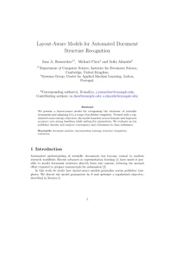
International Journal of Computer Assisted Radiology and Surgery template — frequently asked questions
How do I write a paper in the International Journal of Computer Assisted Radiology and Surgery format?
In DocuGuru you write your manuscript in a normal editor — no LaTeX setup required — and select the International Journal of Computer Assisted Radiology and Surgery template. When you export, DocuGuru compiles the paper into the official Springer Nature format and hands you a submission-ready PDF along with the editable LaTeX source.
What reference style does International Journal of Computer Assisted Radiology and Surgery use?
International Journal of Computer Assisted Radiology and Surgery uses Numbered (Springer Basic) references, shown as numbered [1], [2] markers in the text. DocuGuru formats every in-text citation and the reference list in this exact style automatically. A reference appears like this: 1. Smith A, Jones B, Lee C (2023) A representative article title. International Journal of Computer Assisted Radiology and Surgery 12:45–58
Do I need to know LaTeX to submit to International Journal of Computer Assisted Radiology and Surgery?
No. DocuGuru generates the sn-jnl LaTeX class and compiles the PDF for you in the background, so you get a Springer Nature-ready International Journal of Computer Assisted Radiology and Surgery document without writing any LaTeX. If you do want it, the LaTeX source is included in the export.
Can I import an existing draft into the International Journal of Computer Assisted Radiology and Surgery template?
Yes. Paste or upload your current manuscript — Word, LaTeX, Markdown, or plain text — and DocuGuru reflows it into the International Journal of Computer Assisted Radiology and Surgery format with correct headings, figures, tables, and numbered citations.
Who publishes International Journal of Computer Assisted Radiology and Surgery?
International Journal of Computer Assisted Radiology and Surgery is a multidisciplinary journal published by Springer Nature. DocuGuru's International Journal of Computer Assisted Radiology and Surgery template matches Springer Nature's official submission format.
Can I export a submission-ready International Journal of Computer Assisted Radiology and Surgery PDF?
Yes — DocuGuru produces a PDF built with the official International Journal of Computer Assisted Radiology and Surgery template (the sn-jnl class) that is ready to submit to Springer Nature, together with the matching LaTeX source files.
How much does the International Journal of Computer Assisted Radiology and Surgery template cost?
You can start writing in the International Journal of Computer Assisted Radiology and Surgery template for free. Exporting the final submission-ready International Journal of Computer Assisted Radiology and Surgery PDF and LaTeX source is part of DocuGuru's paid plans — see the app for current pricing.