Elsevier

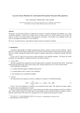
Physics and Imaging in Radiation Oncology Template

Write in a clean editor, then format for Physics and Imaging in Radiation Oncology in one click — DocuGuru applies the official Elsevier template with numbered references and exports a submission-ready PDF plus the editable LaTeX source. Free to start.

About the Physics and Imaging in Radiation Oncology format

Physics and Imaging in Radiation Oncology is a peer-reviewed journal published by Elsevier, covering Advanced Radiotherapy Techniques, Radiation Therapy and Dosimetry, Prostate Cancer Diagnosis and Treatment.

PublisherElsevier
Reference styleNumbered (Elsevier)
Numbered — [1], [2] in the text
[1] A. Smith, B. Jones, C. Lee, A representative article title, Physics and Imaging in Radiation Oncology 12 (2023) 45–58.

Formats any DOI in Physics and Imaging in Radiation Oncology style. No sign-up.

Publishes research inAdvanced Radiotherapy Techniques Radiation Therapy and Dosimetry Prostate Cancer Diagnosis and Treatment Medical Imaging Techniques and Applications Radiomics and Machine Learning in Medical Imaging
ISSN2405-6316
Citation impact (2-yr)1.82
h-index39
i10-index299
Total citations9,318
Article processing charge$1,611
Open accessYes
Top institutions publishing hereAarhus University Hospital
Journal websitewww.journals.elsevier.com
You getA submission-ready PDF and the editable LaTeX source — ready to submit.

Papers published in Physics and Imaging in Radiation Oncology per year

1
2009
1
2012
1
2016
30
2017
65
2018
54
2019
76
2020
76
2021
105
2022
122
2023
161
2024
208
2025

Citation impact of Physics and Imaging in Radiation Oncology by publication year

0
2009
0
2012
0
2016
542
2017
1.2K
2018
1.2K
2019
1.8K
2020
1.3K
2021
1.3K
2022
1K
2023
639
2024
235
2025

Citations each year’s papers have accumulated so far — the most recent years are still building up.

Most-cited papers in Physics and Imaging in Radiation Oncology

Clinical implementation of artificial intelligence-driven cone-beam computed tomography-guided online adaptive radiotherapy in the pelvic region

Patrik Sibolt, Lina Andersson, L. Calmels et al. · 18 Dec 2020

Background and purpose Studies have demonstrated the potential of online adaptive radiotherapy (oART). However, routine use has been limited due to resource demanding solutions. This study reports on experiences with oART in the pelvic region using a novel cone-beam computed tomography (CBCT)-based, artificial intelligence (AI)-driven solution. Material and methods Automated pre-treatment planning for thirty-nine pelvic…

Evaluation of measures for assessing time-saving of automatic organ-at-risk segmentation in radiotherapy

Femke Vaassen, Colien Hazelaar, Ana Vaniqui et al. · 17 Dec 2019

BACKGROUND AND PURPOSE: In radiotherapy, automatic organ-at-risk segmentation algorithms allow faster delineation times, but clinically relevant contour evaluation remains challenging. Commonly used measures to assess automatic contours, such as volumetric Dice Similarity Coefficient (DSC) or Hausdorff distance, have shown to be good measures for geometric similarity, but do not always correlate with clinical applicability of…

Clinical implementation of magnetic resonance imaging guided adaptive radiotherapy for localized prostate cancer

S. Tetar, A. Bruynzeel, Frank J. Lagerwaard et al. · 1 Jan 2019

BACKGROUND AND PURPOSE: Magnetic resonance-guided radiation therapy (MRgRT) has recently become available in clinical practice and is expected to expand significantly in coming years. MRgRT offers marker-less continuous imaging during treatment delivery, use of small clinical target volume (CTV) to planning target volume (PTV) margins, and finally the option to perform daily plan re-optimization. MATERIALS…

Deep learning-based auto-segmentation of targets and organs-at-risk for magnetic resonance imaging only planning of prostate radiotherapy

Sharif Elguindi, Michael J. Zeléfsky, Jue Jiang et al. · 1 Oct 2019

BACKGROUND AND PURPOSE: Magnetic resonance (MR) only radiation therapy for prostate treatment provides superior contrast for defining targets and organs-at-risk (OARs). This study aims to develop a deep learning model to leverage this advantage to automate the contouring process. MATERIALS AND METHODS: Six structures (bladder, rectum, urethra, penile bulb, rectal spacer, prostate and seminal vesicles)…

In vivo dosimetry in external beam photon radiotherapy: Requirements and future directions for research, development, and clinical practice

I. Olaciregui-Ruiz, Sam Beddar, Peter B. Greer et al. · 1 Jul 2020

EPID dosimetry vendors, in collaboration with users across multiple institutions, are requested to improve the understanding and reduce the uncertainties of the system and to help in the determination of optimal action limits for error detection. Finally, the report recommends that automation of all aspects of IVD is needed to help facilitate clinical adoption, including…

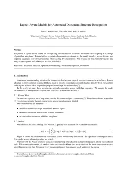
Physics and Imaging in Radiation Oncology template — frequently asked questions

How do I write a paper in the Physics and Imaging in Radiation Oncology format?
In DocuGuru you write your manuscript in a normal editor — no LaTeX setup required — and select the Physics and Imaging in Radiation Oncology template. When you export, DocuGuru compiles the paper into the official Elsevier format and hands you a submission-ready PDF along with the editable LaTeX source.
What reference style does Physics and Imaging in Radiation Oncology use?
Physics and Imaging in Radiation Oncology uses Numbered (Elsevier) references, shown as numbered [1], [2] markers in the text. DocuGuru formats every in-text citation and the reference list in this exact style automatically. A reference appears like this: [1] A. Smith, B. Jones, C. Lee, A representative article title, Physics and Imaging in Radiation Oncology 12 (2023) 45–58.
Do I need to know LaTeX to submit to Physics and Imaging in Radiation Oncology?
No. DocuGuru generates the elsarticle LaTeX class and compiles the PDF for you in the background, so you get a Elsevier-ready Physics and Imaging in Radiation Oncology document without writing any LaTeX. If you do want it, the LaTeX source is included in the export.
Can I import an existing draft into the Physics and Imaging in Radiation Oncology template?
Yes. Paste or upload your current manuscript — Word, LaTeX, Markdown, or plain text — and DocuGuru reflows it into the Physics and Imaging in Radiation Oncology format with correct headings, figures, tables, and numbered citations.
Who publishes Physics and Imaging in Radiation Oncology?
Physics and Imaging in Radiation Oncology is a multidisciplinary journal published by Elsevier. DocuGuru's Physics and Imaging in Radiation Oncology template matches Elsevier's official submission format.
Can I export a submission-ready Physics and Imaging in Radiation Oncology PDF?
Yes — DocuGuru produces a PDF built with the official Physics and Imaging in Radiation Oncology template (the elsarticle class) that is ready to submit to Elsevier, together with the matching LaTeX source files.
How much does the Physics and Imaging in Radiation Oncology template cost?
You can start writing in the Physics and Imaging in Radiation Oncology template for free. Exporting the final submission-ready Physics and Imaging in Radiation Oncology PDF and LaTeX source is part of DocuGuru's paid plans — see the app for current pricing.
Use the Physics and Imaging in Radiation Oncology template