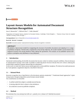
About the Imaging & Microscopy format
Imaging & Microscopy is a peer-reviewed journal published by Wiley, covering Advanced Electron Microscopy Techniques and Applications, Electron and X-Ray Spectroscopy Techniques, Advanced Fluorescence Microscopy Techniques.
| Publisher | Wiley |
|---|
| Reference style | Author–year (Chicago) Author–year — (Smith, 2023) in the text
Smith, Ada, Ben Jones, and Cara Lee. 2023. "A Representative Article Title." Imaging & Microscopy 12 (3): 45–58.
Formats any DOI in Imaging & Microscopy style. No sign-up.
|
|---|
| Publishes research in | Advanced Electron Microscopy Techniques and Applications Electron and X-Ray Spectroscopy Techniques Advanced Fluorescence Microscopy Techniques Cell Image Analysis Techniques Force Microscopy Techniques and Applications |
|---|
| ISSN | 1439-4243 |
|---|
| h-index | 9 |
|---|
| i10-index | 7 |
|---|
| Total citations | 538 |
|---|
| Top institutions publishing here | Carl Zeiss (Germany) |
|---|
| You get | A submission-ready PDF and the editable LaTeX source — ready to submit. |
|---|
Papers published in Imaging & Microscopy per year
Citation impact of Imaging & Microscopy by publication year
Citations each year’s papers have accumulated so far — the most recent years are still building up.
Most-cited papers in Imaging & Microscopy
Tomographic Phase Microscopy
Wonshik Choi, Christopher Fang‐Yen, Seungeun Oh et al. · 1 Mar 2008
In visualizing transparent biological cells and tissues, the phase contrast microscope and its related techniques have been a cornerstone of nearly every cell biology laboratory. However, phase contrast methods are inherently qualitative and lack in 3-D imaging capability. We introduce a novel tomographic microscopy for quantitative three-dimensional mapping of refractive index in live cells and…
Microscopy and Microanalysis 2009
Dave J. Smith · 30 Oct 2009
Huygens Remote Manager
Aaron Ponti, Patrick Schwarb, Asheesh Gulati et al. · 1 Jun 2007
Nowadays, deconvolution in cell and tissue imaging has matured into a standard restoration technique that is accessible to large fraction of the microscopy community thanks to steadily improving algorithms. Still, deconvolution is often the ratelimiting step in the analysis of the acquired data, even at today's computer performance. Here, we present the Huygens Remote Manager,…
Digital In-line Holographic Microscopy
H. J. Kreuzer, M. H. Jericho · 1 Jun 2007
Three-dimensional imaging with micron resolution and simultaneous tracking of many (hundreds) objects (bubbles, beads, algae, bacteria etc.) is now possible in-situ not only with a benchtop microscope, but also remotely in such environments as the deep ocean with a submersible version of the instrument. The instrument is very simple in its hardware requirements and an…
Total Internal Reflection Fluorescence (TIRF) Microscopy
Simon Denham, Deborah Cutchey · 1 May 2009
TIRF microscopy is a technique for imaging the surface of cells that utilizes a thin field of illumination to image fluorophores on or near to the coverslip. TIRF is a highly sensitive widefield camera-based technique providing an excellent signal-to-noise ratio compared with epi-fluorescence or confocal imaging methods. TIRF can be used alone or in combination…