Cambridge University Press

Biological Imaging Template

Write in a clean editor, then format for Biological Imaging in one click — DocuGuru applies the official Cambridge University Press template with numbered references and exports a submission-ready PDF plus the editable LaTeX source. Free to start.

About the Biological Imaging format

Biological Imaging is a peer-reviewed journal published by Cambridge University Press, covering Cell Image Analysis Techniques, Advanced Fluorescence Microscopy Techniques, Advanced Electron Microscopy Techniques and Applications.

PublisherCambridge University Press
Reference styleNumbered
Numbered — [1], [2] in the text
[1] A. Smith, B. Jones, and C. Lee, A representative article title, Biological Imaging 12 (2023) 45–58.

Formats any DOI in the closest standard style — Biological Imaging has no published style definition, so this is an approximation. No sign-up.

Publishes research inCell Image Analysis Techniques Advanced Fluorescence Microscopy Techniques Advanced Electron Microscopy Techniques and Applications Single-cell and spatial transcriptomics Electron and X-Ray Spectroscopy Techniques
ISSN2633-903X
Citation impact (2-yr)4.87
h-index12
i10-index14
Total citations400
Top institutions publishing hereCentre National de la Recherche Scientifique
Journal websitewww.cambridge.org
You getA submission-ready PDF and the editable LaTeX source — ready to submit.

Papers published in Biological Imaging per year

4
2021
8
2022
26
2023
15
2024

Citation impact of Biological Imaging by publication year

92
2021
66
2022
161
2023
68
2024

Citations each year’s papers have accumulated so far — the most recent years are still building up.

Most-cited papers in Biological Imaging

Automated detection and staging of malaria parasites from cytological smears using convolutional neural networks

Mira S. Davidson, Clare Andradi-Brown, Sabrina Yahiya et al. · 1 Jan 2021

Microscopic examination of blood smears remains the gold standard for laboratory inspection and diagnosis of malaria. Smear inspection is, however, time-consuming and dependent on trained microscopists with results varying in accuracy. We sought to develop an automated image analysis method to improve accuracy and standardization of smear inspection that retains capacity for expert confirmation and…

2020 BioImage Analysis Survey: Community experiences and needs for the future

Nasim Jamali, Ellen T. A. Dobson, Kevin W. Eliceiri et al. · 11 Nov 2021

In this paper, we summarize a global survey of 484 participants of the imaging community, conducted in 2020 through the NIH Center for Open BioImage Analysis (COBA). This 23-question survey covered experience with image analysis, scientific background and demographics, and views and requests from different members of the imaging community. Through open-ended questions we asked…

<i>VistoSeg</i> : Processing utilities for high-resolution images for spatially resolved transcriptomics data

Madhavi Tippani, Heena R. Divecha, Joseph Catallini et al. · 1 Jan 2023

Abstract Spatially resolved transcriptomics (SRT) is a growing field that links gene expression to anatomical context. SRT approaches that use next-generation sequencing (NGS) combine RNA sequencing with histological or fluorescent imaging to generate spatial maps of gene expression in intact tissue sections. These technologies directly couple gene expression measurements with high-resolution histological or immunofluorescent images…

FLUTE: A Python GUI for interactive phasor analysis of FLIM data

Dale Gottlieb, Bahar Asadipour, Polina Kostina et al. · 1 Jan 2023

Fluorescence lifetime imaging microscopy (FLIM) is a powerful technique used to probe the local environment of fluorophores. The fit-free phasor approach to FLIM data is increasingly being used due to its ease of interpretation. To date, no open-source graphical user interface (GUI) for phasor analysis of FLIM data is available in Python, thus limiting the…

eHooke: A tool for automated image analysis of spherical bacteria based on cell cycle progression

Bruno M. Saraiva, Ludwig Krippahl, Sérgio R. Filipe et al. · 1 Jan 2021

Abstract Fluorescence microscopy is a critical tool for cell biology studies on bacterial cell division and morphogenesis. Because the analysis of fluorescence microscopy images evolved beyond initial qualitative studies, numerous images analysis tools were developed to extract quantitative parameters on cell morphology and organization. To understand cellular processes required for bacterial growth and division, it…

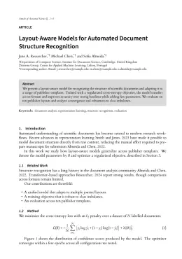
Biological Imaging template — frequently asked questions

How do I write a paper in the Biological Imaging format?
In DocuGuru you write your manuscript in a normal editor — no LaTeX setup required — and select the Biological Imaging template. When you export, DocuGuru compiles the paper into the official Cambridge University Press format and hands you a submission-ready PDF along with the editable LaTeX source.
What reference style does Biological Imaging use?
Biological Imaging uses Numbered references, shown as numbered [1], [2] markers in the text. DocuGuru formats every in-text citation and the reference list in this exact style automatically. A reference appears like this: [1] A. Smith, B. Jones, and C. Lee, A representative article title, Biological Imaging 12 (2023) 45–58.
Do I need to know LaTeX to submit to Biological Imaging?
No. DocuGuru generates the null LaTeX class and compiles the PDF for you in the background, so you get a Cambridge University Press-ready Biological Imaging document without writing any LaTeX. If you do want it, the LaTeX source is included in the export.
Can I import an existing draft into the Biological Imaging template?
Yes. Paste or upload your current manuscript — Word, LaTeX, Markdown, or plain text — and DocuGuru reflows it into the Biological Imaging format with correct headings, figures, tables, and numbered citations.
Who publishes Biological Imaging?
Biological Imaging is a multidisciplinary journal published by Cambridge University Press. DocuGuru's Biological Imaging template matches Cambridge University Press's official submission format.
Can I export a submission-ready Biological Imaging PDF?
Yes — DocuGuru produces a PDF built with the official Biological Imaging template (the null class) that is ready to submit to Cambridge University Press, together with the matching LaTeX source files.
How much does the Biological Imaging template cost?
You can start writing in the Biological Imaging template for free. Exporting the final submission-ready Biological Imaging PDF and LaTeX source is part of DocuGuru's paid plans — see the app for current pricing.
Use the Biological Imaging template